How titanium rods are implanted during surgery
In modern medical fields such as spinal surgery, dental implants, and orthopedic repair, titanium rods, as a high-strength, biocompatible implant material, have become a core component for supporting bone structure and promoting osseointegration. From precise fixation of cervical fractures to mechanical reconstruction of lumbar spondylolisthesis, from alveolar bone anchoring of dental implants to lesion support for spinal tuberculosis, titanium rod implantation technology directly determines surgical success and the quality of patient recovery.

Precise Preoperative Planning: Positioning and Instrument Preparation
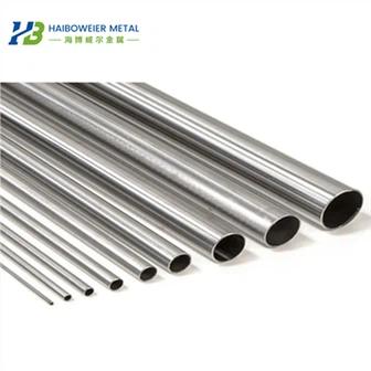
Titanium rod implantation requires anatomical positioning and personalized design. During cervical lateral mass screw-rod fixation, the surgeon uses X-rays or CT scans to determine the anatomical parameters of the pedicles, such as pedicle width, depth, and inclination angle, to select a titanium rod with a diameter of 3.2mm and a length of 120mm or 240mm. For patients with lumbar spondylolisthesis, the distance between the injured vertebra and the adjacent vertebrae must be measured before surgery, and the titanium rod must be pre-bent to match the physiological spinal curvature to avoid postoperative stress concentration and fracture.
The completeness of the surgical instruments is crucial for a successful implantation. Taking thoracic and lumbar fracture internal fixation as an example, the instrument kit must include specialized tools such as a drill sleeve, an adjustable cancellous bone tap, a depth gauge, a rod bender, and a rod holder. The rod bender's bending force must be adjusted based on the material properties of the titanium rod: pure titanium rods are highly ductile and require gradual bending; titanium alloy rods are highly rigid and require complete shaping in one go. Preoperatively, instrument compatibility must be verified through a simulated surgery, such as using a 3D-printed model to test the connection stability between the titanium rod and the pedicle screw.
Intraoperative Stepwise Implantation: From Bone Bed Preparation to Mechanical Locking
Bone Bed Preparation: Drilling and Tapping
The stability of the titanium rod depends on the mechanical lock at the bone-implant interface. During cervical spine surgery, surgeons use a 2.5mm diameter cancellous bone drill bit and drill along the facet articular surface at a 25° angle, maintaining a depth of 18-22mm to avoid penetrating the anterior vertebral cortex. For patients with osteoporosis, self-tapping titanium rods are used instead, as their surface threads enhance bone grip. The tapping process requires strict matching of thread parameters. For example, the pitch of the "stepped" thread must perfectly match the titanium rod thread to prevent micro-motion during insertion that can lead to bone resorption.
Titanium Rod Implantation: Dynamic Adjustment and Minimally Invasive Techniques
In traditional open surgery, titanium rods are manually inserted using a rod holder. For example, in lumbar fusion surgery, the surgeon first places the pre-bent titanium rod into the U-shaped grooves of the pedicle screws on both sides of the injured vertebra. After restoring the vertebral height with a distractor, the nuts are tightened one by one. Minimally invasive techniques utilize percutaneous cannulae for implantation. For example, in percutaneous pedicle fixation, after the titanium rod is inserted through the cannula, its position is confirmed using a bidirectional tracking method. If the minimally invasive cannula moves synchronously with the titanium rod's swing, the rod is securely positioned within the screw slot.
Mechanical Locking: Multi-Level Fixation and Anti-Loosening Design
The ultimate stability of the titanium rod relies on a multi-level locking mechanism. During spinal tuberculosis surgery, the surgeon first secures the rod cap with an elastic nut holder, then applies 50-80N of axial pressure with a persuader to ensure a rigid connection between the titanium rod and the pedicle screw. For cervical spine surgery, an internal locking screw provides additional anti-rotational stability. Its stepped thread design increases locking strength by 30% compared to traditional screws, effectively preventing postoperative rod loosening.
Postoperative Verification and Long-Term Management
Imaging Verification: From 2D to 3D Accurate Assessment
Postoperatively, multimodal imaging with X-rays, CT, and MRI is required to verify the position of the titanium rod. For example, after lumbar spondylolisthesis surgery, sagittal CT reconstructions can clearly demonstrate the relative position of the titanium rod and pedicle. If rod misalignment exceeds 2mm, a secondary surgical adjustment is required. For dental implant titanium rods, micro-CT can quantify the bone-implant contact (BIC). When the BIC is less than 50%, surface coating techniques (such as hydroxyapatite spraying) are indicated to enhance osseointegration.
Functional Rehabilitation: Mechanical Loading and Biofeedback
After titanium rod implantation, appropriate mechanical loading can promote bone remodeling. Following thoracolumbar fracture surgery, patients should gradually increase weight bearing while protected by a brace, transitioning from an initial 20% body weight to full weight bearing to stimulate bone formation around the titanium rod. For patients undergoing cervical spine surgery, electromyography (EMG) monitoring of neck muscle activity is necessary to prevent degeneration of adjacent segments due to excessive titanium rod rigidity.
With advances in materials science and digital medicine, titanium rod implantation technology is evolving towards intelligent and personalized approaches. For example, 3D-printed titanium rods can be customized with a porous structure tailored to the patient's anatomy. Designed with pore sizes of 500-800μm, they promote bone ingrowth and increase bone integrity to over 70%. Furthermore, the introduction of navigation systems and robotic-assisted technology has enabled titanium rod implantation to achieve submillimeter precision, significantly reducing the risk of neurological injury.